


The best tools for diagnosing conditions and assessing the patient’s health status
Examinations performed at our hospital laboratories are characterised by high quality and provide an indispensable source for making accurate medical diagnoses. We offer our patients access to comprehensive ultrasound diagnostics, cardiology diagnostics as well as full laboratory diagnostics.
Cytological examination
Cytology is a diagnostic method used in the prevention and detection of detection of precancerous lesions and cancer of the cervix; the examination is also performed in order to check the results of conservative and surgical treatment of cervical ectropion. Cytology also makes it possible to diagnose infection with HPV (human papillomavirus) which linked to cancer growth. The examination consists of inserting a speculum into the vagina and collecting a sample of cells from the surface of the cervix using a small brush with flexible filaments. The examination is performed on a gynaecology couch by a gynaecologist or midwife; it is completely painless and takes just a few minutes.
Our doctors performing diagnostic tests
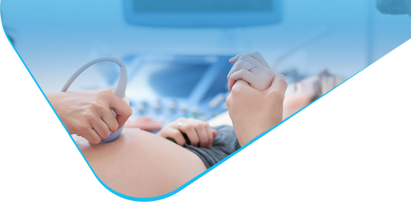
Ultrasound examination
Ultrasound examination is the basic minimally invasive diagnostic method. The examination makes it possible to precisely evaluate the shape, size and depth of the organ concerned. Ultrasonography is also used in prenatal examinations due to the fact that the procedure is completely safe for pregnant women and does not affect the foetus. With modern apparatus we can obtain a reliable three-dimensional image of the organ (3D or 4D). Ultrasound can be used repeatedly on the same person and the examination enables early detection of pathological lesions in internal organs.
Paediatrics:
- Ultrasound of the hip joins (ultrasound of the hips)
- Transfontanellar ultrasound
- Ultrasound of the abdomen
- Ultrasound of the inguinal canals and scrotum
- Ultrasound of the thyroid, salivary glands and lymph nodes
- Ultrasound of the lesser pelvis
Gynaecology:
- Gynaecological ultrasound examinations
- Ultrasound of the breasts
- Cycle monitoring
- Pregnancy ultrasound 3D and 4D
Cardiology:
- Ultrasound of the veins and arteries of upper limbs
- Ultrasound of the veins and arteries of lower limbs
- Ultrasound of the heart (echocardiography)
Endocrinology:
- Ultrasound of the thyroid
- Ultrasound of the parathyroid glands
- Ultrasound of the adrenal glands
- Ultrasound of the salivary glands
- Ultrasound of a goitre
Nephrology, Urology:
- Ultrasound of the kidneys
- Ultrasound of the prostate
- Ultrasound of the urethra
- Ultrasound of the urinary system
- Ultrasound of the testicles
- Ultrasound of the scrotum
Orthopaedics:
- Ultrasound of joints
- Ultrasound of tendons
- Ultrasound of muscles
- Ultrasound of ligaments
Surgery:
- Ultrasound of the veins and arteries of upper limbs
- Ultrasound of the veins and arteries of lower limbs
- Ultrasound of the carotid veins and arteries
- Doppler ultrasound
- Ultrasound of subcutaneous tissue lumps
- Ultrasound of the lymph nodes
- Ultrasound of the abdomen
Preparation for examination
Examination of the abdominal organs:
- the examination should be performed on an empty stomach (you should not eat or drink anything for at least 5 hours beforehand)
- you should not chew gum or smoke before the examination
- patients on medication should take their medicines according to the doctor’s recommendations; not later, however, than 1 hour prior to the examination
- avoid gas-causing foods and drinks, fizzy drinks, yogurts, etc. in the afternoon/evening on the day before the examination
- take Espumisan or a medicine which has a similar effect 24 hours before the examination
Examination of the lesser pelvis (gynaecological, transabdominal, of the urinary bladder and kidneys):
- it is performed with a full bladder so you should drink approx. 1-1.5L still water before the examination. Do not urinate afterwards.
Examination of the prostate:
- for transrectal examination, you should have a bowel movement beforehand (enema is recommended)
- for transabdominal examination, no special preparation is required
Ultrasound of the breasts:
- the examination should best be performed during the first half of the cycle (between day 5 and day 10 of the menstrual cycle)
Vascular Doppler ultrasound:
- patients on medication should take their medicines according to the doctor’s recommendations and wash them down with some still water; not later, however, than 1 hour prior to the examination
- for examination of the kidney artery, aorta, portal vein or hip joints, you should have an empty stomach 6 hours before the examination
Our doctors performing diagnostic tests
Urodynamic testing
Urodynamic testing together with uroflowmetry is a specialist examination that makes it possible to check a lot of voiding parameters (flow rate, urine volume and pressure) and functions of the urinary bladder and the urethra. It is typically performed as a diagnostic examination for urinary incontinence, voiding problems or problems with residual urine. The examination enables assessment of fitness for a procedure aimed at eliminating the ailments. The examination takes approx. 40 minutes and is not painful.
Our doctors performing diagnostic tests
Cystoscopy
Cystoscopy is endoscopy of the urinary bladder, which is applied in diagnostics and treatment of diseases of the urinary system. The procedure consists in direct imaging of the urethra, urinary bladder neck and the urinary bladder mucosa. The physician will insert the cystoscope into the urethra and then up into the bladder in order to fill it up with a liquid. The cystoscope is a kind of endoscope with optical fibres attached to it, which allows the physician to evaluate the status of the urinary organs and collect material for examination, if necessary.
The most common indications for performing this procedure are:
- chronic diseases of the urinary system – especially pain resistant to other therapies;
- recurrent infections of the urinary system;
- irritation of the urinary system, haematuria;
- suspicion of bladder cancer or suspicion of a urinary fistula.
The procedure is performed on a day basis, with local anaesthesia, and the patient can leave the place on their own immediately after the surgery.
Our doctors performing diagnostic tests
Laboratory diagnostics
At the hospital there is a collection point, which means that both elective and urgent examinations can be performed. Most of the tests are performed using one blood sample and the results are usually available already the following day. The types of test performed:
- haematology
- coagulology
- biochemistry
- immunochemistry
- analytics
- serology
- microbiology
Cardiology diagnostics
- ECG– i.e. resting electrocardiography is a popular minimally invasive examination of the heart. An indication for performing the examination may be suspicion of heart dysfunction, i.e. abnormality in the rhythm of the heart (arrhythmia). The examination makes it possible to evaluate the heart rhythm and rate; it also enables detection of myocardial damage in patients who had or are having heart attack (myocardial infarction).
- ECHOCARDIOGRAPHY – is an examination of the structures and movements of the heart by means of an ultrasound apparatus. The examination makes it possible to evaluate the anatomy of the heart, the function of its valves and heart muscle contractility. It is especially recommended to patients with a suspicion of a heart disease.
- STRESS ECG – CARDIAC STRESS TEST – is a minimally invasive examination based on which the physician can evaluate the heart function under increased physical activity. The examination is performed in order to establish an accurate diagnosis for a person with a suspicion of an abnormality in the function of the heart muscle. It makes it possible to establish the prognosis of patients with previous history of myocardial infarction or after coronary artery surgery.
What does prostate fusion biopsy entail?
The objective of the prostate fusion biopsy is to detect prostate cancer; it is performed to confirm a suspected prostate carcinoma. It consists in collecting a tissue sample from areas assessed in an MRI as suspicious, and subsequently in the histopathological examination of the tissue. The examination result is produced in the 3D technology, through the fusion of multi-parametric magnetic resonance imaging (mpMRI) and transrectal ultrasound (TRUS) in real time. The image received eliminates potential deformations resulting from the movement of soft tissues during the MRI and the biopsy procedure performed at the same time. Currently, the fusion biopsy allows for the confirmation of prostate cancer with the highest degree of probability, and its performance reduces necessity of any subsequent biopsies to the minimum.
As the first healthcare facility in the Małopolska region, Szpital na Klinach has the most advanced cutting-edge prostate fusion biopsy system by Koelis Trinity.
What does the examination involve?
The fusion biopsy combines the ultrasound and the MRI. Before collecting a sample, the doctor performs the MRI which allows for pinpointing prostate boundaries and potential cancer lesions. Subsequently, by way of the fusion of images, resonance imaging is transferred in real time to an ultrasound device, which enables the doctor to collect, with great precision, a sample from the relevant area with a potential cancer lesion. During the examination, at least 12 biopsy cores are collected; therefore, sampling from the entire prostate gland, including areas difficult to access, is performed.
The examination is preceded by the doctor entering an anaesthetic gel into the anus, followed by an ultrasound transducer (TRUS). The next step involves collecting samples from the prostate with the use of a biopsy needle under the control of ultrasound imaging. The examination always takes place in a hospital environment, under general or local anaesthesia, with total hospitalisation time of approx. 2.5 hours. The procedure allows for the detection of tumours of 2-3 mm.
Fusion biopsy – benefits
- high cancer detection effectiveness – thanks to the 3D visualisation of the location of each biopsy area, which minimises the risk of overlooking any cancer lesion;
- significant improvement in detecting aggressive lesions in the prostate gland through the flexible fusion of MRI and ultrasound images;
- examination precision – allows for the significantly greater precision of targeting small tumours even of 2-3 mm;
- the patient’s comfort during the examination – the fusion biopsy can be performed under general anaesthesia;
- short hospitalisation time – approx. 5 hours;
- the fusion biopsy reduces necessity of any further biopsies.
To whom is the fusion biopsy dedicated?
The fusion biopsy is a procedure for men applicable in the following cases:
- since 2015, in accordance with a recommendation of the European Association of Urology, each subsequent prostate biopsy should be performed based on the MRI and irregularities detected in such a way;
- the first prostate biopsy is considered – since 2019, the European Association of Urology recommends that each patient in the case of whom the first prostate biopsy is considered should first undergo the MRI of prostate. The resonance is performed precisely with a view to conducting the fusion biopsy;
- increased PSA concentration;
- detected irregularities in rectal palpation (e.g. palpable nodules);
- 4K SCORE or SelectMDx examination results give rise to a suspicion of clinically relevant prostate cancer;
- PCA3 test positive result;
- cells of improper structure have been detected in an earlier biopsy even though it has not shown any neoplastic cells;
- in an mpMRI, suspicious changes have been detected.
Preparation for the biopsy
The fusion biopsy requires proper preparation of the patient. The patient should undergo the examination fasting; further recommendations involve a bowel movement on the examination day and taking an antibiotic. The attending practitioner should always decide about a kind of the antibiotic. Additionally, before the fusion biopsy the patient should inform the doctor of any medications taken (in particular blood thinners), accompanying diseases, a pacemaker and heart valves, if any, and allergies to any drugs. A decision not to take any blood thinners or a decision concerning their dosage is always taken by the doctor. On the examination day, the patient should have the current results of the following tests: APTT, INR, CBC and urinalysis.
Complications after the biopsy
After the prostate biopsy, the patient can experience mild pain and burning in the urethra; however, this symptom should not be a cause for concern and does not require medical intervention. Additionally, the patient can observe blood and clots in his urine, ejaculate and stool, which however should stop on their own after approximately four weeks after the biopsy. Alarming symptoms that require immediate consultation with a urologist are: urinary retention, massive, unstoppable bleeding from the urinary tract or rectum, nausea and vomiting as well as temperature above 38°C, which can point to an infection of the urogenital tract.
How to make an appointment for the prostate fusion biopsy at Szpital na Klinach?
As the first healthcare facility in the Małopolska region, Szpital na Klinach in Krakow has the most advanced cutting-edge prostate fusion biopsy system by Koelis Trinity. The biopsy is performed by: Anna Katarzyna Czech, MD, Tomasz Wiatr, MD and Mateusz Mokrzyś, MD. In order to make an appointment for the examination, please contact our hospital reception staff at the phone number 12 267 40 64 or by e-mail [email protected].


